When a person presents symptoms such as weakness, loss of appetite and weight loss associated with repeated infections or bleeding problems, especially if these appear suddenly, they should go to the doctor to rule out a possible serious origin of these symptoms, such as a leukemia. The diagnosis of ALL should be made by a hematologist, and it is important that it occurs early and completely, determining the type of ALL to properly guide treatment. The presence of ALL is confirmed through various blood, bone marrow, and other diagnostic procedures including:
Physical exam to check general signs of health, signs of disease, lumps, or anything else that seems abnormal. The medical history of the patient’s illnesses and previous treatments are also taken.
Blood test, which will allow us to appreciate the increase in white blood cells.
Bone marrow biopsy, which will allow us to see a lymphoblast ratio greater than 30%.
Genetic and biochemical tests, which will allow determining the ALL subtype.
Imaging tests such as ultrasound, nuclear magnetic resonance or computerized axial tomography, which will make it possible to assess the involvement of other organs.
Blood test and blood cell count
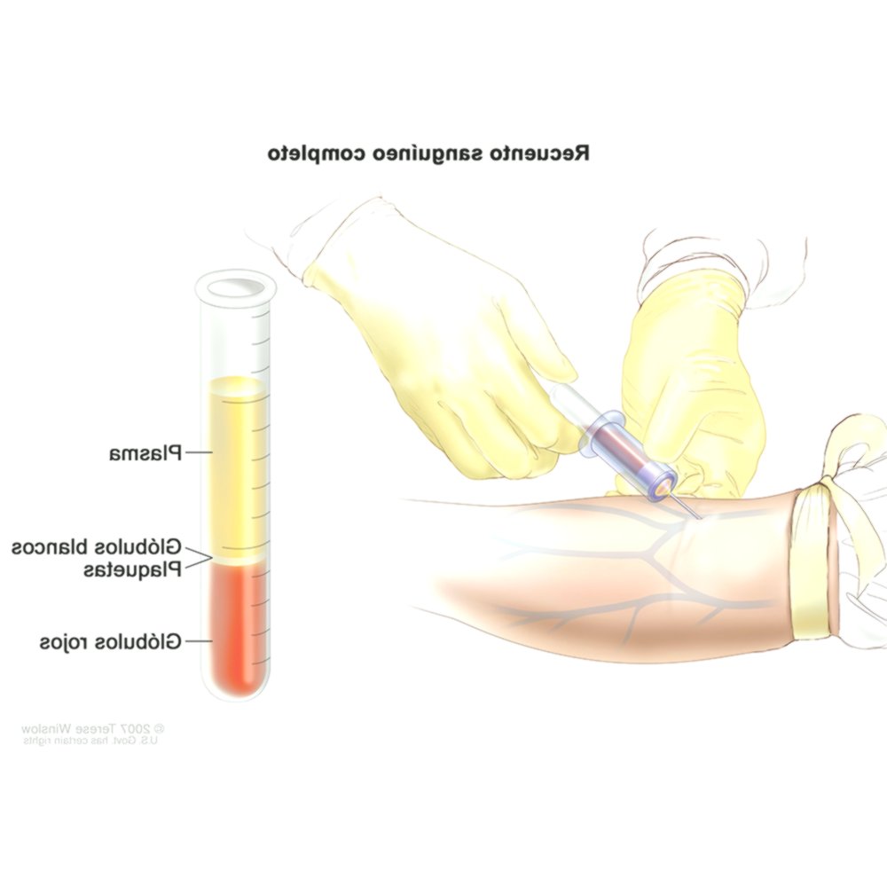
The doctor will order a test called a complete blood count (complete blood count). The blood is drawn in tubes and sent to a laboratory. In this procedure, a small amount of blood is removed from the patient’s arm (sample) with a needle and the following is analyzed:
Number of red blood cells and platelets
Quantity and type of white blood cells
Amount of hemoglobin (the protein that carries oxygen) in the red blood cells
The portion of the sample of red blood cells
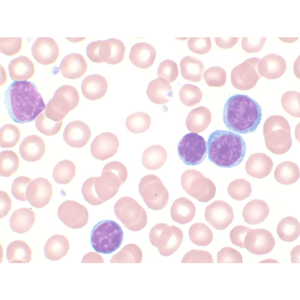
This test measures the number of red blood cells, white blood cells, and platelets. Patients with acute lymphoblastic leukemia generally have fewer red blood cells and platelets than expected. A blood smear may also be done. In this procedure, cells are stained and analyzed under a microscope. A person with acute lymphoblastic leukemia often has too many lymphoblasts in their blood. Lymphoblasts are immature (young) cells that do not function like normal mature cells.Blood smear from a child with acute lymphoid leukemia The image above is not representative of a lymphoblastic leukemia, it makes one think more of a chronic lymphatic leukemia, I suggest that they change it. The blood smear sample can also be used for cytogenetic analysis and immunophenotyping, which are explained below.
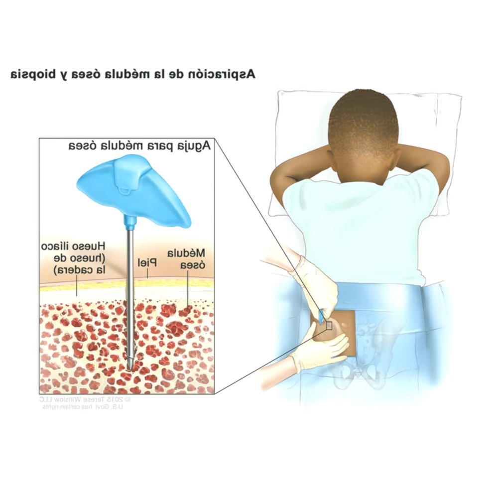
Bone marrow tests
Other tests will be done to ensure that the diagnosis of acute lymphoblastic leukemia is correct. These tests routinely include bone marrow aspiration and bone marrow biopsy, which allow bone marrow samples to be studied under a microscope. Bone marrow aspiration: A sample of cells is removed from the bone marrow. Bone marrow biopsy: A small piece of bone filled with bone marrow is removed. Both bone marrow tests are done with a special needle that is usually inserted into the hip bone or sternum. Before starting the procedure, the patient is given medicine to numb the part of the body from which the cell sample will be taken. Some patients receive a medication that makes them sleepy during this procedure and others remain awake. The cell sample is usually drawn from the patient’s hip bone (iliac bone). A specialist looks at the bone marrow and bone samples under a microscope to check for signs of cancer. Blood and bone marrow tests can be done in the doctor’s office or in a hospital. Aspiration and bone marrow biopsy are almost always done in the same office. Blood and bone marrow tests are not only performed at diagnosis, but are also repeated during and after treatment, to verify that the treatment is working and that it destroys acute lymphoblastic leukemia cells.
Cytogenetic analysis
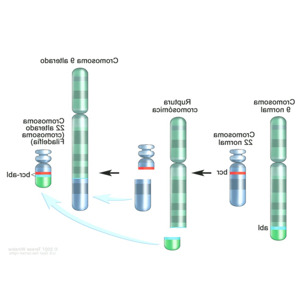
All cells in the body have chromosomes2 that contain the genes. Genes provide instructions that tell each cell what to do. The test called cytogenetic analysis is used to examine the chromosomes of blast cells for acute lymphoblastic leukemia. This test, which has been carried out on the blood or bone marrow tissue sample, has made it possible to identify a large number of recurrent chromosonic disorders and is considered essential for the classification of patients in the different treatment lines. Some of the alterations identified through cytogenetic analysis are associated with very specific biological and clinical characteristics, which have been used as diagnostic and prognostic markers and have contributed to the definition of risk groups. For example, in the ALL with the Philadelphia chromosome. The alteration that results when parts of chromosome 9 and chromosome 22 are exchanged is called the Philadelphia chromosome.Philadelphia Chromosome : A section of chromosome 9 and a section of chromosome 22 break and exchange places. The bcr-abl gene is formed on chromosome 22 where it joins the section of chromosome 9. The altered chromosome 22 is called the Philadelphia chromosome.ImmunophenotypingThis procedure is used to diagnose specific types of leukemia by comparing cancer cells with normal cells of the immune system. Immunophenotyping also serves to separate cells into different groups according to the antigens and proteins (markers) they have on their cell surface from a blood or bone marrow cell, in order to find out if they are lymphocytes or myeloid cells, and if acute lymphoblastic leukemia cells are B cells or T cells. Most people have acute B cell lymphoblastic leukemia.Fluorescence in situ hybridization (FISH)This test is another way to examine chromosomes and genes. It uses special fluorescent dyes that only adhere to specific genes or parts of particular chromosomes. The FISH test can find most of the chromosomal changes (such as translocations) that are visible under a microscope in conventional cytogenetic tests, as well as some changes that are too small to see with the usual cytogenetic test.The FISH test can be used on routine blood and bone marrow samples. Because cells do not have to divide for this test, it can also be used to examine cells from other tissues, such as lymph node samples. This test is very accurate and can usually provide results in a short time, just a few days.Polymerase chain reaction (PCR)The polymerase chain reaction is a highly sensitive DNA test that can also find some certain chromosomal changes so small that they cannot be seen under a microscope, even if the sample has very few leukemic cells. Like FISH, it was used to find particular genetic changes and not to examine chromosomes in general.In the case of ALL, it is frequently used to identify the presence of the gene produced by the Philadelphia chromosome.If the leukemic cells have a particular genetic change (or chromosome), PCR can be used after treatment to try to find small numbers of leukemic cells that may not be visible under a microscope.Lymph node biopsyRemoval of a lymph node or part of a node is often done to help diagnose lymphomas, but may be necessary, sometimes in leukemias, although the diagnosis can usually be made by testing the blood and bone marrow .In this procedure, a surgeon cuts the skin to remove all or part of a lymph node. If the node is close to the surface of the skin, this is a simple operation that can often be done under local anesthesia, but if the node is inside the chest or abdomen, general anesthesia is used to keep you asleep during the biopsy. .When a lymph node is completely removed, it is called a lymph node excision biopsy. If part of a lymph node is removed, it is called a lymph node incision biopsy.Lumbar punctureLumbar puncture is a procedure used to take a sample of cerebrospinal fluid. It is done by placing a needle between two bones in the spine into the cerebrospinal fluid that surrounds the spinal cord, and a sample of the fluid is removed. The cerebrospinal fluid sample will be studied for leukemic cells that may have spread to the central nervous system.Occasionally, a lumbar puncture is performed after the diagnosis of leukemia to determine if the leukemic cells have spread to the brain and spinal cord. Intrathecal chemotherapy3 is given after removal of a fluid sample to treat leukemic cells that may have spread to the brain or spinal cord.IMAGING STUDIESImaging tests use sound waves, x-rays, magnetic fields, or radioactive particles to take pictures of the inside of the body. Because leukemia usually does not form tumors, imaging tests are not as helpful as they are for other cancers.Imaging studies can be done in ALL patients, but they are most often done to detect infections or other problems, not for the leukemia itself. In some cases they can be done to help determine the extent of the disease, if you think it may have spread by affecting organs other than the bone marrow and blood.Chest x-rayX-ray of the organs and bones inside the chest. An x-ray is a type of energy beam that can go through the body and be reflected on a plate that shows an image of the inside of the body. Chest radiography is a simple, quick and very useful technique.Computed axial tomographyComputed tomography is a type of radiological study that produces a detailed, cross-sectional image of your body. Unlike a regular x-ray, CT scans can show detail in soft tissues (such as internal organs).This study can help detect if any of your lymph nodes or organs are enlarged. It is generally not needed to diagnose ALL, but it can be done if your doctor suspects that the leukemia may be infiltrating other organs such as your spleen.Instead of taking a single image like conventional x-rays do, a CT scanner takes many images while rotating around the body. Then a computer combines these photographs into detailed images of the part of the body being studied.Before the scan, you may be asked to take a contrast solution and / or receive an intravenous (IV) injection of a contrast material that helps to better map abnormal areas of the body. You may need an intravenous (IV) line to inject the contrast material. IV injection of the contrast dye may cause a flushing or warm feeling on the face or other areas of the body. Some people are allergic and have hives or rarely more serious reactions such as difficulty breathing and a drop in blood pressure. Be sure to tell the doctor if you are allergic to anything or if you have ever had a reaction to any contrast material used for x-rays.The CT scan used for this study consists of a ring similar to a large thread, with a narrow stretcher located in the central opening. You will have to lie still on the table while the test is done. CT scans take longer than conventional radiographs, and you may feel a little confined by the ring while the images are being taken.In some cases, CT can be used to accurately guide a biopsy needle to the suspected abnormality, such as an abscess. For this procedure, called a computed tomography-guided needle biopsy, you stay on the CT table as a radiologist moves a biopsy needle through the skin and into the mass. CT scans are repeated until the needle is inside the mass. Then, a biopsy sample is extracted and will be studied and analyzed.Sometimes a study is done that combines CT with positron emission tomography (PET / CT scan). For positron emission tomography (PET), a form of radioactive sugar (known as fluordeoxyglucose or FDG) is injected into the blood. The amount of radioactivity used is low. Because cancer cells in the body grow rapidly, they absorb large amounts of sugar. A special camera can then create an image of the areas of radioactivity in the body. PET / CT allows the physician to compare the areas of greatest radioactivity on PET with the appearance of that area on CT. This is not often done in ALL patients.Magnetic resonance imagingLike computed tomography, magnetic resonance imaging (MRI) provides detailed images of the soft tissues of the body. However, MRI uses powerful radio waves and magnets instead of X-rays. The energy of the radio waves is absorbed by the body and then released in a pattern formed by the type of tissue in the body and by certain diseases. A computer translates the pattern into a highly detailed image of body parts. Gadolinium, a contrast material, is often injected into a vein before the study is done to better show the details. This contrast material is different from that used for CT.MRI exams are also very useful for examining the brain and spinal cord.Magnetic resonance imaging takes longer than CT scans, it can take over an hour. During the study, you may lie in a narrow tube, which can be uncomfortable and confining for some people. Another option is the new MRI machines that are more open. The MRI machine produces a loud, hammering hum that can be uncomfortable. Hearing aids or ear plugs are offered in some places to help block this noise.Ultrasound (ultrasound)Ultrasound uses sound waves and their echoes to produce an image of the internal organs or masses. For this study, a small, microphone-like instrument called a transducer (which is first lubricated with gel) is usually placed on the skin. This instrument emits sound waves and collects the echo that bounces off the organs. A computer converts the echoes into an image that is displayed on a computer screen.It can be used to look at lymph nodes near the body’s surface, or to look at swollen organs inside your abdomen, such as the kidneys, liver, and spleen.This test is easy to perform and does not use radiation. For most ultrasounds, you simply lie on a stretcher, and a specialist moves the transducer to the part of your body that is being studied.Gallium scan and bone scanThese studies are used infrequently for ALL, but can be helpful if you have bone pain that may be due to either a bone infection or cancer involving the bones.For these studies, the doctor or nurse injects a slightly radioactive chemical into the bloodstream. The chemical builds up in areas of cancer or infection that can be seen with a special type of camera. The images from these studies look like “radioactive spots” on the body, but they don’t provide much detail. If an area is illuminated in the study, other imaging studies, such as X-rays, CT, or MRI, may be done to get a more detailed picture of the area. If there is a possibility of leukemia, this may need to be confirmed by biopsy of the area.
CONDITIONS OF USE OF THE SERVICE
The information provided by this means cannot, in any way, replace a direct health care service, nor should it be used for the purpose of establishing a diagnosis, or choosing a treatment in particular cases. In this service no recommendation will be made, explicitly or implicitly, about drugs, techniques, products, etc … that will be cited for informational purposes only. The use of this service is carried out under the exclusive responsibility of the users
We use cookies to ensure that we give you the best experience on our website. Continued use of this site indicates that you accept this policy. Read more. Accept